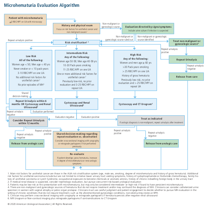
Microscopic hematuria algorithm, from AUA Guidelines
Assessing hematuria risks
Epidemiology
- 3+ RBCs on microscopic UA warrant assessment and potential hematuria workup
- 1 urinalysis with RBCs is all that is required for a hematuria workup
- 6.5% healthy patients have microscopic hematuria on screening tests
- 2.5% (0.7-4.3%) patients with asymptomatic microscopic hematuria have underlying malignant cause
- 33-67% patient do not have diagnosible cause of hematuria (positional, physical activity, recent intercourse)
Risk factors
- Smoking: increases risk of bladder cancer
- Radiation: XRT cystitis, secondary malignancy, fistula
- Chemotherapy: cyclophosphamide can lead to hemorrhagic cystitis
- UTI: hematuria may be a presenting symptom
- Chronic catheter: irritation or development of SCC bladder cancer
- Chemical exposure: benzenes, aromatic amines increases risk of bladder cancer
- Hx stones, BPH, trauma: benign causes of hematuria
- Dysuria: in absence of UTI, may be presenting symptom of carcinoma in situ
- Hx cancer: either GU-specific or syndromic (Lynch, VHL)
Hematuria workup
Risk Groups
Risk |
Needs all criteria? |
Age |
Smoking (packyears) |
# RBC (UA x1) |
Urothelial cancer risk factors* |
Prior hematuria |
Probability malignant cause |
Recommendations |
Low |
Yes |
Women < 50 Men < 40 |
< 10 |
3-10 |
None |
No |
0.2-0.5% |
Repeat UA in 6mo, OR... Cysto + renal US |
Intermediate |
No |
Women 50-59 Men 40-59 |
10-30 |
11-25 |
1+ |
Persistent 3-10 RBC |
1.3-1.6% |
Cysto + renal US |
High |
No |
60+ |
> 30 |
> 25 |
- |
Gross |
10.8-11.1% |
Cysto + CTU |
*Risk factors include increased age, male, prior smoking, irritative voiding symptoms, prior pelvic radiation, prior cyclophosphamide, history urothelial cancer or Lynch syndrome, prior occupational exposures, and chronic indwelling foreign body
Considerations
- Hematuria in setting of UTI does not warrant workup, perform workup if hematuria persists after treatment (wait 3wks-3mo)
- Patients on anticoagulation should undergo same workup
- If benign cause identified and treatment, discuss with patient whether further workup is warranted
- Perform full workup even if nephrologic cause suspected (increased risk for GU cancers in setting of decreased renal function)
- Patients should undergo thorough upper tract imaging if family history RCC or genetic syndrome
Workup components
- Upper tract imaging: CTU, MRU, renal US + retrograde pyelograms
- Renal US: less sensitive for upper tract urothelial cancer but very sensitive for renal neoplasms, but due to rarity the costs outweigh the benefits
- Cystoscopy: perform without enhancement (blue light), preferable as screening test
- If persistent/worsening hematuria after renal US, obtain further imaging
- Cytology: obtain only if persistent microhematuria, irritative voiding symptoms, or risk for CiS
- Labs: consider BMP if nephrologic concerns, PSA if male and > 40yo, can also consider CBC and coags
Follow-up
- Low risk (no workup) and negative repeat UA at 12mo no further workup
- Int/high risk and negative initial workup: repeat UA in 12mo
- Repeat UA negative: no further workup
- Persistent microhematuria: discuss need for further workup immediately or in 3-5yrs
- Worsening hematuria: obtain further workup
Hematuria grading scale Option #1, from Lee 2013
Hematuria grading scale Option #2, from Stout 2021
Glomerular causes of hematuria, from Campbell's
Hematuria management
Initial Evaluation of Gross Hematuria
- Safe to discharge: able to empty bladder (check bladder scan), no blood clots (check urine appearance), not actively bleeding (stable Hgb, non-hypotensive)
- Retention with mild bleeding: likely prostatic origin, attempt regular catheter placement (often results in clear yellow urine, catheter bypasses prostatic bleeding)
- Retention likely secondary to clot burden: likely prior XRT or surgery, place large 3-way Rusch catheter, manually irrigate, start continuous bladder irrigation (CBI)
- Persistent bleeding despite CBI: consider traction, OR clot evacuation, other cause-specific treatments
BPH
- Cause: increased vascularity in hyperplastic tissue secondary to VEGF
- Finasteride: inhibits androgen-stimulated angiogenesis and decreases VEGF expression, 90% symptom improvement, decreased need for surgical intervention, can take 2 weeks to 9 months to take effect
- Refractory hematuria: best managed with surgical outlet procedures
Prostate Cancer
- Usually caused by locally advanced cancer with bladder invasion
- Management: consider ADT, palliative XRT, channel TURP, cystoprostatectomy w/ diversion
Urethrorrhagia
- Trauma: most common non-gender specific cause, maintain catheter for 3-7 days
- Urethritis can be infectious or chemical induced
- Urethral tumors: consider if history urothelial cancer
Upper tract causes
- Presentation: clot colic, anemia, and wormlike clots in urine
- Nephropathies: look for dysmorphic RBCs and casts on UA, systemic symptoms
- Papillary necrosis: sickle cell, NSAID use
- Localized management: may require embolization or partial/total nephrectomy
Random causes of hematuria to rule out
- TB/Schistosomiasis: travel to endemic areas
- ADPKD: family history
- Endometriosis or uterouro fistula: cyclical hematuria during menstrual cycle
- Ureteroiliac fistula: hx vascular surgery, chronic ureteral stents, pelvic XRT
- Arteriovenous malformation: may occur if recent renal procedure, treat w/ embolization
- Nutcracker syndrome: compression of renal vein between aorta and SMA
Hemorrhagic Cystitis
Common Causes
- XRT: seen in up to 5% after pelvic XRT
- Chemotherapy: seen in 2-40% after cyclophosphamide, ifosfamide, resolves in 60-90% with mesna
- Viral (BK polyoma): more common in children or immunocompromised
Medical Management
Drug |
Mechanism |
Dosing |
Considerations |
Needs OR/Anesthesia? |
Contraindications |
Side effects |
Alum Aluminum ammonium sulfate Aluminum potassium sulfate |
Protein precipitation Vasoconstriction |
1% solution (10g/L) run at 200-300mL/hr
|
Success: 45-100% No need for anesthesia Can give with VUR |
No |
None |
Aluminum toxicity |
Amicar Aminocaproic acid |
Inhibits fibrinolysis |
1g/L intravesical 5g PO loading dose + 1g/hr Give for 24hr after hematuria resolves |
Success: up to 92% |
Bladder clots present (causes them to harden) DIC Upper tract bleeding (causes glomerular thrombosis) Risk factors for thrombosis |
Rhabdomyolysis (monitor CPK if used for > 24hr) Hypotension GI effects |
Silver Nitrate |
Chemical coagulation |
0.5-1% instilled for 10-20 minutes Rinse out with saline |
Mix with water (will precipitate in saline) |
Sometimes (if high concentrations) |
Extravasation VUR (need to occlude ureters) Inability to tolerate general/spinal anesthesia |
Bladder scarring Ureteral strictures if VUR |
Formalin |
Cellular protein precipitation |
1-4% solution 300mL or up to bladder capacity hold for 10-15 minutes Irrigate bladder with 1L water/saline |
Success: 80-90% |
Yes |
Hyperbaric oxygen
- Technique: 100% O2 at 2-3 atm, 90 minutes, 30-40 sessions
- Benefits: enhances angiogenesis, vasoconstriction, antibiotic efficacy, neutrophil function
- Absolute contraindications: cisplatin/doxorubicin treatment, untreated pneumothorax, active viral infections
- Relative contraindications: seizure risk, poorly controlled DM, emphysema, optic neuritis, glaucoma, current pregnancy, fever, active malignancy, hx sinus/ear surgery, hx spontaneous pneumothorax, spherocytosis, claustrophobia
- Success for XRT cystitis: 80-90% response rate, but 5yr success only 27%
- Side effects: claustrophobia (20%), otalgia (17%), seizures (rare)
Surgical/procedural interventions
Treatment |
Tips |
Nephrostomy tubes |
Avoids bladder exposure to urokinase, allowing clots to form Can be performed with ureteral coiling |
Internal iliac artery embolization |
Can be performed unilaterally/bilaterally Posterior occlusion results in significant gluteal pain Success: up to 90% |
Cystectomy + Urinary Diversion |
Complications in up to 80% if bladder not removed High risk of complications |
Sterile Pyuria
Common Causes
- Infectious: Tb, STI, Mycoplasma, Ureaplasma, recently treated UTI, Schistosomiasis, Adenovirus, prostatitis, intraurethral HPV, HIV, fungus, Trichomonas
- Weight loss: Tb, pelvic malignancy
- Prior surgery/XRT: pelvic XRT, recent cystoscopy, stents/PCNs/catheters
- Hematuria: stones, ADPKD, renal papillary necrosis, tubulointerstitial nephritis (TIN)
- Pertinent PMHx: SLE, DM, Kawasaki, pregnancy, malignant HTN
- Pertinent medications: NSAIDs, steroids, olsalazine, nitrofurantoin, penicillins, vancomycin, PPI
Workup
- Initial workup (PCP): Tb-PCR, STI screen, renal US, cytology
- Eosinophilia: seen in tubular interstitial nephritis (TIN), Schistosomiasis
- Urologic workup: cystoscopy + CTU if concerned
Different causes of urine color, from Campbell's
Proteinuria algorithm, from Campbell's
Other UA findings
Things that look like hematuria but aren't hematuria
- Myoglobinuria: seen during rhabdo, can cause positive blood on a dipstick but will be negative for RBCs on the microscopic UA
- Pyridium: turns urine bright orange, will cause positive nitrites without leukocyte esterase
- Menstruation: confirm clean catch (check squamous cells)
Non-hematuria findings
- Excess urine on dipstick or holding vertical can alter results
- Phosphaturia: phosphate crystals in alkaline urine, can cause non-infected turbid urine appearance, urine will immediately clear with acidification of sample
- UTI + pH > 7.5: suggests presence of urea-splitting organism
- RBC casts: indicate glomerular source, usually associated with proteinuria
- Proteinuria: always from renal source, may have false negative if albumin is not the primary protein (Tamm-Horsfall, Bence-Jones protein)
- Glucosuria: seen when glucose levels exceed renal reabsorption threshold
- Ketonuria: seen with DKA, pregnancy, starvation, weight loss
- Microscopic examination:: assess for cells, casts, crystals, bacteria, yeast, and parasites
References
- AUA Core Curriculum
- Barocas, Daniel A., et al. "Microhematuria: AUA/SUFU Guideline." The Journal of urology 204.4 (2020): 778-786.
- Boorjian, et al. "Evaluation and management of hematuria." Campbell-Walsh Urology. 12th ed. Philadelphia, PA: Elsevier (2020): 247-259.
- Castle, E., C. Wolter, and M. Woods. "Evaluation of the Urologic Patient: Testing and Imaging." Campbell-Walsh Urology 12 (2020).
- Glen, Peter, Akash Prashar, and Amr Hawary. "Sterile pyuria: a practical management guide." The British Journal of General Practice 66.644 (2016): e225.
- Goonewardene, Sanchia, and Raj Persad. "Sterile pyuria: a forgotten entity." Therapeutic advances in urology 7.5 (2015): 295-298.
- Lee, Joo Yong, et al. "Hematuria grading scale: a new tool for gross hematuria." Urology 82.2 (2013): 284-289.
- Stout, Thomas E., Michael Borofsky, and Ayman Soubra. "A visual scale for improving communication when describing gross hematuria." Urology 148 (2021): 32-36.
- Wieder JA: Pocket Guide to Urology. Sixth Edition. J.Wieder Medical: Oakland, CA, 2021.
- Wise, Gilbert J., and Peter N. Schlegel. "Sterile pyuria." New England Journal of Medicine 372.11 (2015): 1048-1054.